• Skip to primary navigation
  • Skip to main content
  • Skip to primary sidebar
  • Skip to footer

Johns Hopkins Vasculitis Center

Show Search
Hide Search
  • About Our Center
    • Appointments and Directions
    • Meet Our Team
    • Support Our Center
  • What is Vasculitis?
    • Types of Vasculitis
    • Causes of Vasculitis
    • Symptoms of Vasculitis
    • Diagnosing Vasculitis
  • Vasculitis Treatments
    • Prednisone
    • Avacopan (Tavneos®)
    • Apremilast (Otezla®)
    • Azathioprine
    • Colchicine
    • Cyclophosphamide (Cytoxan)
    • Dapsone
    • Supplemental Immunoglobulin (IVIG/SCIG)
    • Leflunomide
    • Mepolizumab (Nucala®)
    • Methotrexate (MTX)
    • Mycophenolate
    • Rituximab
    • Sarilumab (Kevzara®)
    • TNF Inhibitors
    • Tocilizumab (Actemra®)
  • Vasculitis Research
  • Resources
Home / Search for "polyarteritis nodosa"

Search Results for: polyarteritis nodosa

Causes of Vasculitis

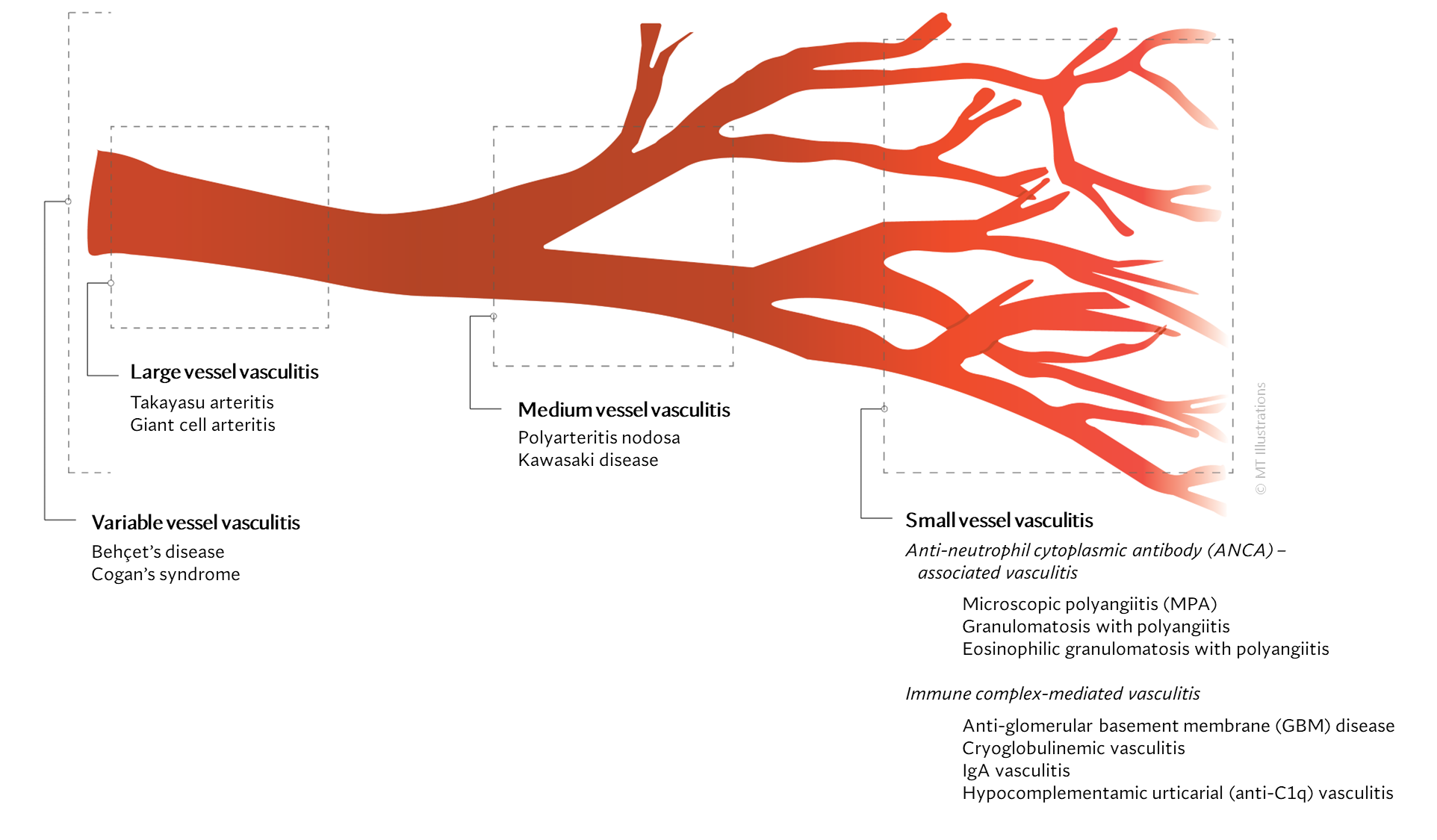
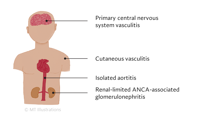
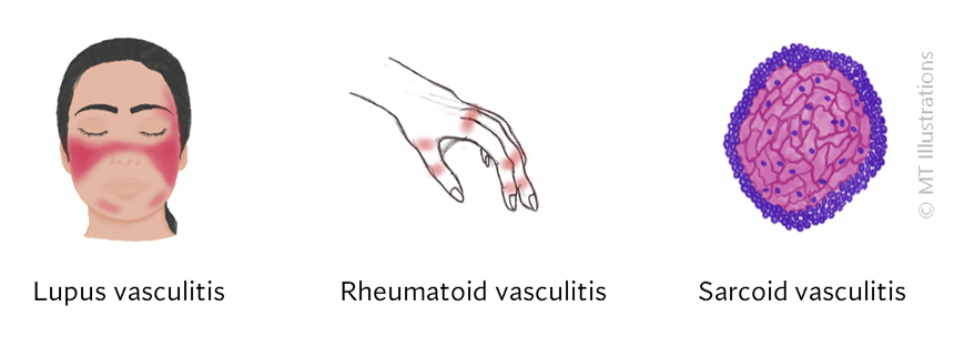
There are many different types of vasculitis, some with different causes than others.

Certain forms of vasculitis that can be due to infection where the microbe directly invades the vessel wall. Syphilis is one example of vasculitis that can be caused by infection in the blood vessel. Treating the infection is the main goal in managing this sort of vasculitis, which is not an autoimmune disease, but rather an infection.

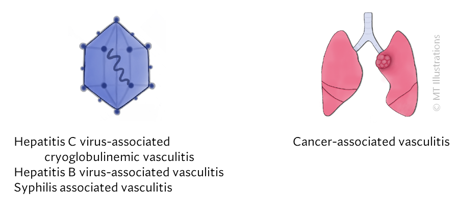
Other infections can provoke the immune system into causing damage in blood vessels. Here, the infection is the trigger, but the immune system is the cause of the vascular damage. Viral hepatitis (B and C) are examples of this sort: some patients with Hepatitis B may develop polyarteritis nodosa, while some patients with Hepatitis C may develop cryoglobulinemic vasculitis.

Other types of vasculitis may be due to an ‘allergic‘-type reaction to medications. For example, certain blood pressure medications (hydralazine) or thyroid medications (propylthiouracil) can trigger ANCA associated vasculitis in some patients. Cocaine is an illicit drug that is linked to vasculitis and vascular damage.

However, the causes of most vasculitides are currently unknown. While we can identify some risk factors (such as older age in giant cell arteritis), we do not know the specific causes of these diseases. These forms of vasculitis of unknown cause are considered autoimmune diseases.

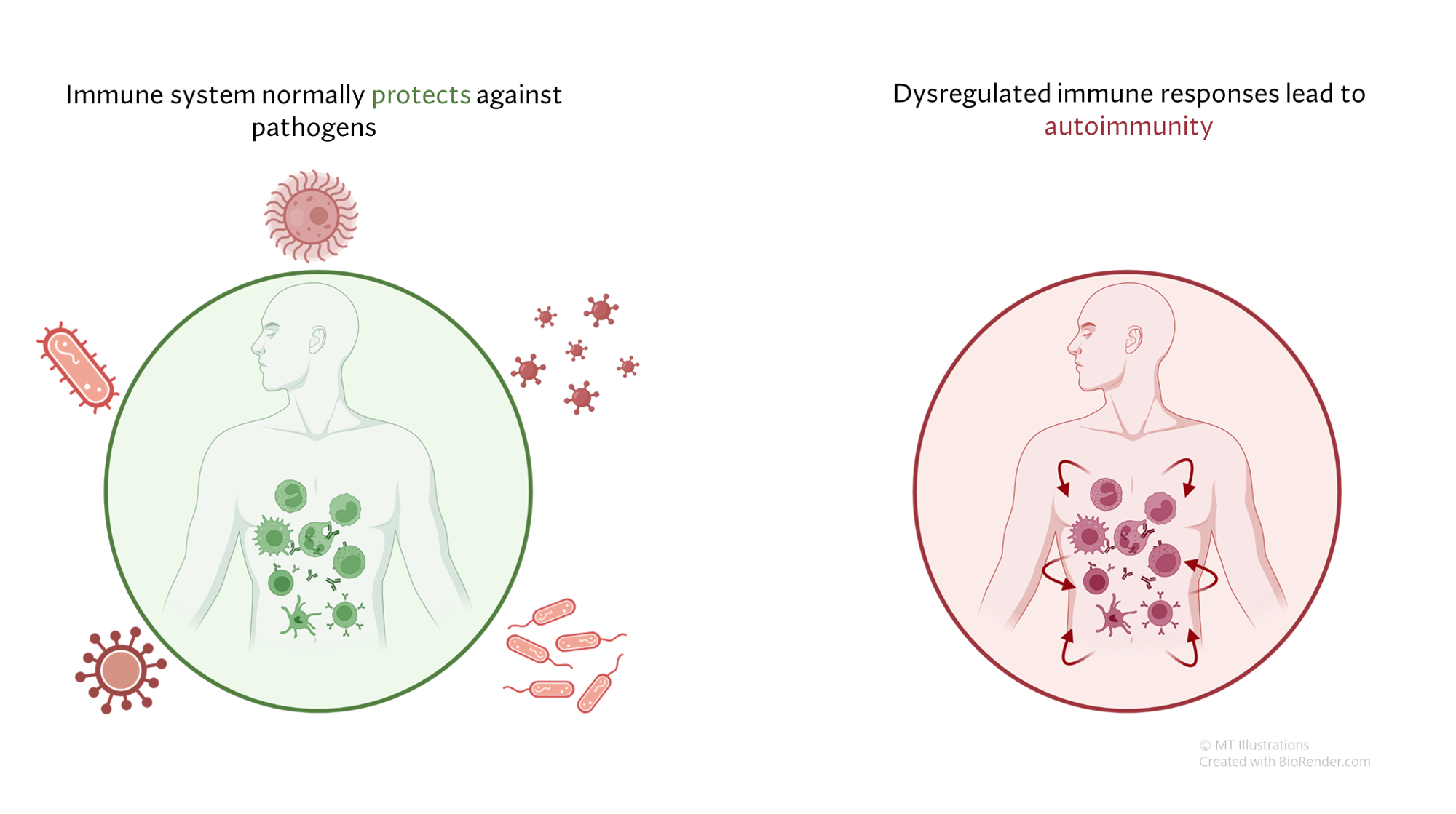
Under normal circumstances, our immune system serves to defend us from infection and other threats, such as cancers. In autoimmune diseases, the immune system generates a response not against a foreign threat, but against normal “self” tissues. This abnormal immune response against “self” tissues can result in a wide array of autoimmune diseases, including relatively common diseases (such as psoriasis or thyroid disease) as well as rare conditions (such as vasculitis).

In most cases, autoimmune diseases are believed to be due to an abnormal immune response that is generated in a susceptible person, and eventually leads to a cycle of ongoing inflammation in otherwise normal tissues where no infection or other identifiable threat is present. Some interaction between the immune system and the environment is thought necessary for this to occur, and a person’s genetic background likely places some individuals at higher risk than others.

A better understanding of the specific causes of these diseases would lead to improved means of diagnosing, treating, and even preventing these conditions. Uncovering the causes of vasculitis is a major goal of vasculitis research.

While we may not know the specific causes of the vasculitidies, we do have a basic understanding of the way that the immune system causes organ damage in these conditions. In all forms of vasculitis, activation of the immune system leads to the deposition of inflammatory cells and proteins in the walls of blood vessels. As this inflammation in blood vessels continues, the vessels become damaged and no longer serve their normal function of delivering blood to the organs that they supply. Consequently, the tissues downstream of these inflamed vessels are starved of oxygen and nutrients needed for normal function. At a basic level, this is a process similar to what occurs in a heart attack or a stroke – but instead of the cholesterol plaque that blocks a coronary artery in a heart attack, the immune system is responsible for blockage of blood vessels in vasculitis.

All information contained within the Johns Hopkins Vasculitis Center website is intended for educational purposes only. Visitors are encouraged to consult other sources and confirm the information contained within this site. Consumers should never disregard medical advice or delay in seeking it because of something they may have read on this website.

Types of Vasculitis

There are approximately 20 different disorders that are classified as “vasculitis”. “Angiitis” and “Arteritis” are both synonyms for vasculitis, literally meaning “inflammation within blood vessels” or “inflammation in arteries.”  Because there are so many types of vasculitis, the group is sometimes referred to in the plural: vasculitides (pronounced “vas que lit’ i deez”).

There are many different types of diseases that belong to this category. Although the diseases are similar in some ways, they often differ with respect to which organs are affected, which medications are used to treat them, and other characteristics.

In general, vasculitis can be grouped based on the size of the blood vessels that are
mainly affected. However, it is important to keep in mind that any sized blood vessels can be
involved.

Large vessel vasculitis (LVV)
LVV is characterized by inflammation of the largest-sized blood vessels of the body such
as the aorta, major arteries that deliver blood to distant parts of the body. They also include
large veins that deliver blood back to the heart. These are:

  • Takayasu arteritis
  • Giant cell arteritis

Medium vessel vasculitis (MVV)
MVV is inflammation of the medium-sized blood vessels including arterioles and smaller
veins.

  • Polyarteritis nodosa
  • Kawasaki disease

Small vessel vasculitis (SVV)
SVV affects the smallest blood vessels of the body called capillaries and venules. SVV
involves inflammation mediated by autoantibodies (antibodies are molecules that usually coats
bacteria and viruses; however, in autoimmune disease, they bind to cells and proteins normally
found in the body, hence the name, autoantibodies). These antibodies can deposit in small vessels
causing restriction of blood flow to tissue.
They can be of two types:

(1) Anti-neutrophil cytoplasmic antibody (ANCA)-associated vasculitis (AAV)

  • Microscopic polyangiitis (MPA)
  • Granulomatosis with polyangiitis (GPA)
  • Eosinophilic granulomatosis with polyangiitis (EGPA)


(2) Immune complex small vessel vasculitis

  • Anti-glomerular basement membrane
  • Cryoglobulinemic vasculitis
  • IgA vasculitis
  • Hypocomplementamic urticarial vasculitis (anti-C1q vasculitis)

Variable vessel vasculitis (VVV)
This type of vasculitis can involve inflammation of blood vessels of any size.

  • BehÇet’s disease
  • Cogan’s syndrome

Others:

Single-organ vasculitis (SOV):
SOV is inflammation affecting the blood vessels that feed a single organ, such as the brain or skin.

Vasculitis associated with another autoimmune disease:
Vasculitis can occur in conjunction with other autoimmune diseases, such as lupus or sarcoidosis.

Vasculitis associated with other diseases:
Other diseases, such as infections and cancer, can cause systemic vasculitis as well.
Vasculitis as a side effect of drugs:
Certain drugs, such as hydralazine and cocaine, can cause vasculitis.

Vasculitis Frequently Asked Questions

  • What causes vasculitis?
  • What is going to happen to me?
  • Is vasculitis curable?
  • Is vasculitis hereditary?
  • Does diet affect vasculitis?
  • Will my vasculitis return?
  • How should I guard against the occurrence of a disease flare?
  • Why do I have to have bloodwork checked frequently?

What causes vasculitis?

The causes of most forms of vasculitis remain unknown. Infections are strongly suspected of playing a role in in forms such as the association of hepatitis B (a virus) and polyarteritis nodosa, and hepatitis C (another virus) and cryoglobulinemic vasculitis. Bacterial infections have been suspected of playing a possible role in granulomatosis with polyangiitis (GPA, formerly known as Wegener’s) which is the reason that some patients with GPA that is limited to the upper respiratory tract are treated only with an antibiotic, Bactrim (trimethoprim/sulfamethoxazole). A general theory that applies to many types of vasculitis is that the disease results from the occurrence of a particular infection in a person whose genes (and other factors) make him/her susceptible to developing vasculitis.

What is going to happen to me?

The course of vasculitis is often difficult to predict. Some types of vasculitis may occur only once and do not return. Other types are prone to recurrences. For all patients with vasculitis, it is essential to be evaluated by physicians who are experienced in the treatment of these diseases. Vasculitis is treatable, and many patients achieve remissions through treatment. It is important to balance the types of medications necessary to control the disease and the risk of side effects that those medicines often bring. A primary aim of several ongoing new studies in vasculitis is to find drugs that help maintain remission.

Is vasculitis curable?

Most forms of vasculitis are treatable if detected early enough, before substantial organ damage has occurred. While often effective, however, the treatments remain imperfect and require improvement. Further research is needed in all forms of vasculitis. Greater knowledge of these diseases will lead to better treatments and, some day, to cures.

Will my children or other family members get it?

Vasculitis is not contagious. One cannot acquire vasculitis from contact with a vasculitis patient. In addition, despite the fact that genes probably play a role in susceptibility to some forms of vasculitis, it is unusual for vasculitis to occur in more than one member of the same family. Thus, vasculitis is not a heritable disorder. All of these points illustrate the fact that the causes of vasculitis are complex. In all likelihood, patients develop vasculitis because of the simultaneous occurrence of multiple risk factors, most of which remain poorly understood.

Does diet affect vasculitis?

This is one of the most commonly-asked questions by patients with vasculitis. All patients want to do whatever is within their power to help treat their disease. Unfortunately, there is presently no evidence that a person’s diet affects susceptibility to vasculitis, or that consuming or avoiding certain foods or beverages affects the course of the disease. In general, we advocate eating a balanced healthy diet rich in protein and vegetables. Avoidance of excessive empty calories, processed foods, and sugars may be very important, particularly in patients on steroids who are at risk for weight gain.

Will my vasculitis return?

After patients achieve remission from their vasculitis, it is logical for them to wonder if their disease will ever return. The answer, which is often difficult to give with certainty, depends in large part on the patient’s specific type of vasculitis. For example, some types of vasculitis, such as Henoch-Schönlein purpura (HSP) or vasculitis caused by a medication, are often self-limited and resolve on their own. Other forms of vasculitis (e.g., Buerger’s disease, a disease strongly associated with cigarette smoking) resolve with institution of the definitive treatment: smoking cessation.

However,  other forms of vasculitis behave less predictably and never come back in some patients but recur frequently in others. Granulomatosis with polyangiitis (GPA), giant cell arteritis (GCA), Takayasu arteritis, microscopic polyangiitis, and many other types of vasculitis fall into the category of diseases that have periods of quiescence and periods of flare. Disease flares in vasculitis can be mild (rash, minor joint pains) or severe (renal failure, skin ulcers). Flares may occur if medications are discontinued or dosage is lowered. Flare may occur in the context of infection. Often the reason for disease flare is unknown.

At the present time, the ability of doctors to predict who will suffer disease flares and who will maintain in long-term remissions (or be cured) needs refinement. Progress in this area will come through research.

How should I guard against the occurrence of a disease flare?

We believe that several points are worth keeping in mind:

First, the symptoms of flares are usually very similar those experienced at the onset of disease. If headaches signaled the beginning of giant cell arteritis, then the recurrence of headaches may indicate a disease flare. If leg ulcers began as painful red lumps on the leg the first time, then the return of painful red lumps may mean that vasculitis is back. Patients must become experts about their own manifestations of vasculitis so that they can recognize them immediately, consult their doctors, and begin appropriate treatment before serious damage occurs.

Second, we believe that patients truly know and understand their own bodies. It is important to discuss new or changing symptoms with your physicians. Together, patients and physicians can determine if new symptoms truly represent a vasculitis flare or if the cause is something equally as likely (medication side effect, infection, or other common medical issues).

Finally, because vasculitis treatments require careful monitoring by doctors, patients should discuss any changes in treatment with their physicians. Increasing or decreasing medications without consulting a physician may lead to trouble.

Why do I have to have bloodwork checked frequently?

Blood tests are helpful to monitor for the return of vasculitis by keeping a watchful eye on important parameters such as kidney function, liver tests, and markers of inflammation (ESR and CRP). Blood tests are also very important to ensure that medications are not causing any side effects such as liver irritation or low blood counts.

How often should my blood be checked?

This depends on the specific medicine or medicines that you take. Patients on cyclophosphamide (Cytoxan) should have their counts checked every week. Patients on most other kinds of medications used to treat vasculitis (Methotrexate, Azathioprine) usually only need to have their blood work checked monthly. If some laboratory tests are abnormal or nearly so, then more frequent monitoring may be required.

What type of tests do we check?

Regardless of the type of vasculitis and the exact type of medication that a patient takes, similar types of tests are monitored. These tests are:

  1. a complete blood count;
  2. tests of kidney function including a urinalysis; and
  3. liver function tests.

The table below outlines the importance behind checking each of these tests.

Type of TestWhat should be checkedWhy?
Complete Blood Count (“CBC”)
  • White blood cells (WBC)
  • Platelets
  • Hematocrit
  • Low WBC count may lead to infections.
  • Low platelets may cause bleeding.
  • Low hematocrit means insufficient oxygen-carrying capacity of the blood.
Kidney Function
  • Creatinine
  • Blood Urea Nitrogen (BUN)
  • High creatinine and BUN indicate that the kidneys are not performing their blood-cleansing function properly.
Urinalysis
  • Protein Level
  • Red Blood Cells
  • Normal urinalyses have no protein and no blood.
  • The presence of protein and/or blood in the urine may indicate active vasculitis in the kidneys (or damage to the bladder from cyclophosphamide).
Liver Function
  • Albumin
  • Aspartate aminotransferase(AST)
  • Alanine aminotransferase (ALT)
  • Often a good indication of overall health.
  • Elevated AST/ALT levels indicate inflammation in the liver (usually caused by medications).

Buerger’s Disease

  • First Description
  • Who gets Buerger’s Disease (the “typical” patients)?
  • Classic symptoms of Buerger’s Disease
  • What causes Buerger’s Disease?
  • How is Buerger’s Disease diagnosed?
  • Treatment and Course of Buerger’s Disease

First Description

This disease was first reported by Buerger in 1908, who described a disease in which the characteristic pathologic findings — acute inflammation and thrombosis (clotting) of arteries and veins — affected the hands and feet. Another name for Buerger’s Disease is thromboangiitis obliterans.

Who gets Buerger’s Disease (the “typical” patient)?

The classic Buerger’s Disease patient is a young male (e.g., 20–40 years old) who is a heavy cigarette smoker. More recently, however, a higher percentage of women and people over the age of 50 have been recognized to have this disease. Buerger’s disease is most common in the Orient, Southeast Asia, India and the Middle East, but appears to be rare among African–Americans.

Classic symptoms and signs of Buerger’s Disease

The initial symptoms of Buerger’s Disease often include claudication (pain induced by insufficient blood flow during exercise) in the feet and/or hands, or pain in these areas at rest. The pain typically begins in the extremities but may radiate to other (more central) parts of the body. Other signs and symptoms of this disease may include numbness and/or tingling in the limbs and Raynaud’s phenomenon (a condition in which the distal extremities — fingers, toes, hands, feet — turn white upon exposure to cold). Skin ulcerations and gangrene (pictured below) of the digits (fingers and toes) are common in Buerger’s disease. Pain may be very intense in the affected regions.

An angiogram demonstrating lack of blood flow to vessels of the hand (figure below). This decreased blood flow (“ischemia”) led to ulcers of the fingers and severe pain.

An abnormal result from an angiogram of the hand (figure below).

Despite the severity of ischemia (lack of blood flow) to the distal extremities that occurs in Buerger’s, the disease does not involve other organs, unlike many other forms of vasculitis. Even as ulcers and gangrene develop in the digits, organs such as the lung, kidneys, brain, and gastrointestinal (GI) tract remain unaffected. The reasons for the confinement to the extremities and sparing of other organs are not known.

What Causes Buerger’s Disease?

The association of Buerger’s Disease with tobacco use, particularly cigarette smoking, cannot be overemphasized. Most patients with Buerger’s are heavy smokers, but some cases occur in patients who smoke “moderately”; others have been reported in users of smokeless tobacco. It has been postulated that Buerger’s Disease is an “autoimmune” reaction (one in which the body’s immune system attacks the body’s own tissues) triggered by some constituent of tobacco.

Pictured below, are a patient’s fingertips that have developed gangrene. This is a very painful condition which sometimes requires amputation of the affected area.

How is Buerger’s diagnosed?

Buerger’s disease can be mimicked by a wide variety of other diseases that cause diminished blood flow to the extremities. These other disorders must be ruled out with an aggressive evaluation, because their treatments differ substantially from that of Buerger’s Disease (for Buerger’s, there is only one treatment known to be effective: complete smoking cessation — see below).

Diseases with which Buerger’s Disease may be confused include atherosclerosis (build–up of cholesterol plaques in the arteries), endocarditis (an infection of the lining of the heart), other types of vasculitis, severe Raynaud’s phenomenon associated with connective tissue disorders (e.g., lupus or scleroderma), clotting disorders of the blood, and others.

It should be noted that other substances, such as marijuana, have also been associated with a vasculitis similar to Buerger’s or polyarteritis nodosa that should be considered in the differential diagnosis.

Angiograms of the upper and lower extremities can be helpful in making the diagnosis of Buerger’s disease. In the proper clinical setting, certain angiographic findings are diagnostic of Buerger’s. These findings include a “corkscrew” appearance of arteries that result from vascular damage, particularly the arteries in the region of the wrists and ankles. Angiograms may also show occlusions (blockages) or stenoses (narrowings) in multiple areas of both the arms and legs.

Pictured below on the left is a normal angiogram. On the right, is an abnormal angiogram of an arm demonstrating the classic “corkscrew” appearance of arteries to the hand. The changes are particularly apparent in the blood vessels in the lower right hand portion of the picture (the ulnar artery distribution).

In order to rule out other forms of vasculitis (by excluding involvement of vascular regions atypical for Buerger’s), it is sometimes necessary to perform angiograms of other body regions (e.g., a mesenteric angiogram).

Skin biopsies of affected extremities are rarely performed because of the frequent concern that a biopsy site near an area poorly perfused with blood will not heal well.

Treatment and Course of Buerger’s

It is essential that patients with Buerger’s disease stop smoking immediately and completely. This is the only treatment known to be effective in Buerger’s disease. Patients who continue to smoke are generally the ones who require amputation of fingers and toes.

Despite the clear presence of inflammation in this disorder, anti-inflammatory agents such as steroids have not been shown to be beneficial. Similarly, strategies of anticoagulation (thinning of the blood with aspirin or other agents to prevent clots) have not proven effective. The only way to prevent the progression of the disease is to abstain from all tobacco products.

Eosinophilic Granulomatosis with Polyangiitis, formerly Churg-Strauss Syndrome (EGPA)

  • First Description
  • Who gets EGPA (the “typical” patients)?
  • Classic symptoms of EGPA
  • What causes EGPA?
  • How is EGPA diagnosed?
  • Treatment and Course of EGPA

First Description

Eosinophilic granulomatosis with polyangiitis (EGP), formerly known as the Churg-Strauss Syndrome , is a systemic vasculitis. This disease was first described in 1951 by Dr. Jacob Churg and Dr. Lotte Strauss as a syndrome consisting of “asthma, eosinophilia [an excessive number of eosinophils in the blood], fever, and accompanying vasculitis of various organ systems”. EGPA shares many of the clinical and pathological features of polyarteritis nodosa (“PAN”, another type of vasculitis) and granulomatosis with polyangiitis (GPA). However, Drs. Churg and Strauss discovered that the presence of granulomas as well as the abundance of eosinophils distinguished this disease from PAN and GPA.

Who gets EGPA (the “typical” patient)?

The typical patient with EGPA is a middle aged individual with a history of new-onset or newly-worsened asthma. The distribution of the disease among males and females is approximately equal.

Classic symptoms and signs of EGPA

Asthma is one of the cardinal features of EGPA. Asthma symptoms may begin long before the onset of vasculitis – e.g., many years before any other symptoms of EGPA arise, and long before the diagnosis of EGPA is made. Other early symptoms/signs include nasal polyps and allergic rhinitis.

The next phase of the disease is often marked by eosinophilia, the finding of an excessive number of eosinophils in the blood or in tissues. An eosinophil is one subtypes of white blood cell. Normally, eosinophils comprise 5% or less of the total white blood cell count. In EGPA, the percentage of eosinophils may reach as high as 60%. In the picture below, the eosinophils are shown by the dark pink stain.

The third phase of the illness is a vasculitis, which involves the skin, lungs, nerves, kidneys, and other organs. Particular mention should be made of the frequent devastating involvement of the nerves (called mononeuritis multiplex), which produces severe tingling, numbess, shooting pains, and severe muscle wasting/power loss in the hands or feet. The list below contains the organs commonly involved by EGPA and the specific disease manifestation(s) in each organ.

  • Nose
  • Lung
  • Skin
  • Kidney
  • Gastrointestinal
  • Heart
  • Nerve

Nose

  • Sinusitis, including allergic rhinitis
  • Nasal polyps

Lung

  • Pulmonary infiltrates (only one-third of all patients)
  • Bleeding into the lungs (occasionally)
  • Diffuse interstitial lung disease (rarely)

Skin

  • Rashes
  • Palpable purpura
  • Nodules (above or below the skin), often at sites of pressure, such as the elbows

Kidney

  • Glomerulonephritis (inflammation in the small units of the kidney that filter blood)
  • Hypertension

Gastrointestinal

  • Lesions (vasculitic) are occasionally found in the GI tract
  • Granuloma sometimes found in spleen

Heart

  • Vasculitis lesions in heart, can lead to congestive heart failure or a heart attack

Nerve

  • Peripheral nerve involvement including pain, numbness, or tingling in extremities (neuropathy/mononeuritis multiplex)

What causes EGPA?

The cause of EGPA is unknown but is probably multi-factorial. Genetics may play a small role in the disease, but EGPA is almost never seen in two members of the same family. Environmental factors such as exposure to industrial solvents may play a role in susceptibility to this disease, but this is largely speculative. Infections may be the inciting event(s), but to date there is no definitive evidence of this.

How is EGPA Diagnosed?

Among all of the vasculitides, asthma is a distinctive feature of EGPA alone. However, not all patients with asthma have vasculitis – only a tiny minority do, in fact. It is the specific combination of symptoms and signs, the pattern of organ involvement, and the presence of certain abnormal blood tests (eosinophilia, in particular) that help the doctor make the diagnosis. In addition to a detailed history and physical examination, blood tests, chest X-rays and other types of imaging studies, nerve conduction tests, and tissue biopsies (e.g., of lung, skin, or nerve) may be performed to help diagnose EGPA.

The following features are consistent with a diagnosis of GPA:

  1. asthma
  2. eosinophilia [>10% on differential WBC count]
  3. mononeuropathy
  4. transient pulmonary infiltrates on chest X-rays
  5. paranasal sinus abnormalities
  6. biopsy containing a blood vessel with extravascular eosinophils.

Treatment and Course of EGPA

EGPA usually responds to prednisone. Initially, high doses of oral prednisone are used in an attempt to get the disease into remission as quickly as possibly (e.g., using oral prednisone 40-60 mg/day). After the first month or so, this high dose of prednisone is gradually tapered down over the ensuing months. Other immunosuppressive drugs, such as azathioprine, cellcept, methotrexate, cyclophosphamide, or rituximab may be used in addition to prednisone. High doses of intravenous steroids (usually methylprednisolone) maybe useful for those patients with severe disease or for those who are unresponsive to the combination of oral prednisone used with other immunosuppressive medications.

Prior to the advent of prednisone, EGPA was often a fatal disease. The majority of patients died from rampant, uncontrolled disease. With present therapy, constitutional symptoms begin to resolve quite quickly, with gradual improvement in cardiac and renal function, as well as improvement in the pain that results from peripheral nerve involvement. The course of therapy can last for 1 to 2 years, although the length and type of treatment depend on the severity of disease and the organs involved. The patient’s response to treatment and the continuation of disease control during lowering of the prednisone dose are the primary determinants of how long therapy is continued. Laboratory monitoring of blood tests is very helpful in gauging the activity of disease. Some of the most useful laboratory tests are the erythrocyte sedimentation rate (ESR) and the eosinophil count.

  • « Go to Previous Page
  • Go to page 1
  • Go to page 2

Primary Sidebar


donate to the Johns Hopkins Vasculitis Center

All information contained within the Johns Hopkins Vasculitis Center website is intended for educational purposes only. Visitors are encouraged to consult other sources and confirm the information contained within this site. Consumers should never disregard medical advice or delay in seeking it because of something they may have read on this website.

Footer

Johns Hopkins Rheumatology

  • Johns Hopkins Rheumatology
  • Johns Hopkins Arthritis Center
  • Johns Hopkins Lupus Center
  • Johns Hopkins Lyme Disease Research Center
  • Johns Hopkins Myositis Center
  • Johns Hopkins Scleroderma Center
  • Johns Hopkins Sjögren’s Syndrome Center

Connect With Us

  • Facebook
  • Twitter
  • YouTube
U.S. News and World Report Rankings Badge

Johns Hopkins Medicine

© 2025 Johns Hopkins Vasculitis Center
Patient Privacy